![]()
Congratulations to B02 and C01 on their successful joint publication in Biomedical Optics Express.
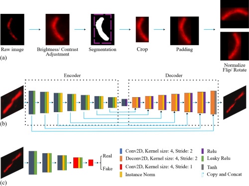
With the growing integration of artificial intelligence in scientific and medical applications, lightweight deep learning models have become increasingly important. These models offer substantial reductions in memory usage and computational time. Given that GPU-based model training and inference contribute significantly to carbon emissions, lightweight architectures with comparable performance to parameter-rich models present a more environmentally friendly alternative. Specifically, we build upon CycleGAN with a fixed-channel lightweight U-Net generator for modality transfer from standard confocal to super-resolution STED and deconvolved STED images, and systematically compare it against Pix2Pix and standard CycleGAN baselines. Obtaining paired datasets in medical imaging and super-resolution microscopy is often infeasible due to the need for additional experiments and the intrinsic complexity of biological sample preparation. To address this, we investigate the performance of lightweight CycleGAN models, demonstrating their ability to achieve high-fidelity modality transfer despite reduced model complexity. We introduce a fixed channel strategy within the U-Net-based generator, in contrast to the traditional channel-doubling approach. This modification significantly reduces the number of trainable parameters from 41.8 million to approximately 9 thousand, while achieving comparable or slightly improved performance. We explore the utility of GAN models as a qualitative marker for assessing experimental and labeling quality. When trained on high-quality microscopy images, the GAN implicitly learns the characteristics of optimal imaging. Deviations between GAN-generated outputs trained on high-quality data and low-quality experimental images can highlight potential issues such as photobleaching, experimental artifacts, or inaccurate labeling. In this way, the model can support qualitative assessment of experimental consistency and image fidelity in fluorescence microscopy workflows.