![]()
C03: Deciphering virus-membrane interactions with advanced optical microscopy and machine

Projects of the CRC 1768
C03: Deciphering virus-membrane interactions with advanced optical microscopy and machine
A critical step in virus infection is the initial uptake of viruses into host cells. Interestingly, and perhaps counterintuitively, a particular virus may use different modes of crossing the plasma membrane, either in the same cell or depending on cell type. Even for members of the same virus family, different modes of uptake have been observed. The main uptake pathways include the use of membrane fusion or endocytosis. A number of endocytic mechanisms have been discovered and these depend on multiple factors, such as cell type and surface receptors, as well as cell membrane properties. Importantly, the uptake path usually determines the fate and infectivity of a virus. Thus, predicting the mode of entry is important for choosing and optimising preventative or impeding measures of infections.
However, determining an uptake mechanism is challenging due to multiple factors involving assay design and observation technology. An accurate investigation requires an adapted experimental design for each virus, receptor and host cell, analysing binding, uptake, routing, and the infection cycle of cellular and virus proteins. At the same time, direct microscopic imaging of the viruses and other molecules involved in the uptake into living cells should employ as high a temporal and spatial resolution as possible, since every entry mode displays specific spatiotemporal dynamics of virus and molecular movements at the cell and virus surface.
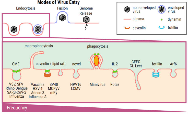
Sketch of different uptake mechanisms, ranging from various endocytosis pathways to membrane fusion to the injection of genetic material (as for bacteriophages).
Finally, data analysis needs to be tailored to correlate a “microscopic signature” of entry events with fusion signatures and particular endocytic mechanisms, which would then make it possible to identify the mode(s) of entry of an emerging virus. Unfortunately, the realisation of high-resolution microscope experiments of virus uptake is usually too complex to accomplish a straightforward and quick prediction of entry modes of newly emerging viruses. This is, however, within the overall aim of this CRC VirusREvolution, which is to set up tools to quickly react to potential pandemic threats. We therefore propose to realise a fast and facile tool to predict the mode(s) of virus entry from standard low-resolution live-cell microscopy data. To achieve this, we will combine high spatiotemporal
resolution microscopy data with methods from Machine Learning (ML) / Artificial Intelligence (AI) for fast, objective, and quantitative analysis to establish a tool that allows correlation with the standard microscopy data of lower spatiotemporal resolution. This shall enable the characterisation and differentiation of the membrane interactions of virtually anyvirus, usingwidelyusedstandardandlesscomplexmicroscopyapproaches,suchaswidefieldorconfocal microscopy. We will employ a machine learning approach that builds on the experience gained from working with a variety of viruses. Thus, the performance of our tool will gradually improve as more and more virus data becomes available. Our experimental and computational tools will contribute to answering the research questions of the CRC VirusREvolution, such as revealing mechanisms of virus entry and tackling the goals of the description and prediction of the infectivity of viruses and their hosts.
Project Overview
Central to our project plan is the realisation of a fast and facile tool to initially predict the mode(s) of virus entry from standard low-resolution live-cell microscopy data. Underlying this approach is the idea that virus entry modes can be differentiated by virus particle kinetics and signal intensity profiles prior to and during fusion or endocytic uptake, and can be linked to mechanistic information. These parameters depend on (1) receptor engagement and its mobility in the membrane, and (2) critically, the mode of crossing the plasma membrane. While (1) may vary, (2) is limited to distinct patterns. We propose to elucidate these patterns and link them to mechanistic information on virus entry. To this end, we will record correlative live-cell virus-entry data from low-resolution standard and high-resolution SRM fluorescence microscopy, and employ computational parameters to enable the extraction of in-depth information from standard techniques. To achieve this, we will combine standard and SRM microscopy data and employ machine learning methods for fast, objective, and quantitative analysis of entry modes of virtually any virus with standard microscopy data only, Fig. C03.4. We will utilise a machine learning approach that will gradually improve as more and more virus data becomes available. Overall, we will realise (a) the direct visualisation of virus membrane interactions, (b) tracking of virus entry events, and (c) prediction of the mode of entry for viruses. The aim is that the improved understanding of the virus entry mechanism will open new therapeutic developments. We combine expertise in employing standard microscopy to virus entry analysis (Schelhaas lab), development of SRM and its application towards membrane characterisation and virus cycling (Eggeling lab), and unique knowledge of data analysis, including ML algorithms to disclose various cell-biological events, including pathogen-cell interactions (Figge lab).
- Tool to be developed: Optical microscopy-based tools to provide fast and facile predictions of the mode of entry of emerging pathogens.
- Tool to be developed: Optical microscopy protocols for simultaneous recording and correlation of standard and high resolution data of virus-membrane interactions.
- Tool to be developed: Machine learning-supported workflow in JIPipe for fast, objective, and quantitative image analysis and classification of virus-cell interactions.
Hypothesis enabled by the proposed tool: The prediction of the mode of virus uptake is important for choosing preventive measures of infections by emerging pathogens, and adaptation of optical microscopy and data analysis will provide the required tools.
Overarching CRC goals: Our project C03 couples high-spatiotemporal single-virus imaging with a machine-learning pipeline that infers entry modalities from standard widefield/confocal time-lapse data, enabling rapid, scalable annotation of membrane-interaction signatures across diverse viruses (G1, G3). Trained on growing consortium datasets, the classifier provides calibrated predictions of fusion vs. endocytic uptake routes for emerging pathogens, supporting early triage of infectivity-modifying interventions (G4).
Work Packages (WP):
- WP 1: Optimised visualisation of virus-membrane interactions (Eggeling/Schelhaas)
- WP 2: Fast prediction of virus entry modes through correlation of techniques (Eggeling/Figge/Schelhaas)
- WP 3: Machine-Learning-supported image analysis of virus-membrane interaction (Figge/Eggeling/SchelhaaS)
Team Members


Dr. Ziliang Zhao
Postdoc
Dr. Ruman Gerst
Postdoc
Ivan Avilov
Postdoc
Dr. Dhruv Khatri
Postdoc
2025
Brinkert, Pia; Krebs, Lena; Ventayol, Pilar Samperio; Greune, Lilo; Bannach, Carina; Amakiri, Cynthia; Bucher, Delia; Kollasser, Jana; Dersch, Petra; Boulant, Steeve; others,
The actin nucleation promoting factor WASH facilitates clathrin-independent endocytosis of human papillomaviruses Journal Article
In: EMBO reports, vol. 26, no. 22, pp. 5533, 2025.
@article{brinkert2025actin,

title = {The actin nucleation promoting factor WASH facilitates clathrin-independent endocytosis of human papillomaviruses},

author = {Pia Brinkert and Lena Krebs and Pilar Samperio Ventayol and Lilo Greune and Carina Bannach and Cynthia Amakiri and Delia Bucher and Jana Kollasser and Petra Dersch and Steeve Boulant and others},

year  = {2025},

date = {2025-01-01},

urldate = {2025-01-01},

journal = {EMBO reports},

volume = {26},

number = {22},

pages = {5533},

keywords = {},

pubstate = {published},

tppubtype = {article}

}
2024
Dasgupta, Anindita; Koerfer, Agnes; Kokot, Boštjan; Urbančič, Iztok; Eggeling, Christian; Carravilla, Pablo
Effects and avoidance of photoconversion-induced artifacts in confocal and STED microscopy Journal Article
In: Nature Methods, vol. 21, no. 7, pp. 1171–1174, 2024.
@article{dasgupta2024effects,

title = {Effects and avoidance of photoconversion-induced artifacts in confocal and STED microscopy},

author = {Anindita Dasgupta and Agnes Koerfer and Boštjan Kokot and Iztok Urbančič and Christian Eggeling and Pablo Carravilla},

year  = {2024},

date = {2024-01-01},

urldate = {2024-01-01},

journal = {Nature Methods},

volume = {21},

number = {7},

pages = {1171–1174},

publisher = {Nature Publishing Group US New York},

keywords = {},

pubstate = {published},

tppubtype = {article}

}
2023
Rizzato, Matteo; Mao, Fuxiang; Chardon, Florian; Lai, Kun-Yi; Villalonga-Planells, Ruth; Drexler, Hannes CA; Pesenti, Marion E; Fiskin, Mert; Roos, Nora; King, Kelly M; others,
Master mitotic kinases regulate viral genome delivery during papillomavirus cell entry Journal Article
In: Nature Communications, vol. 14, no. 1, pp. 355, 2023.
@article{rizzato2023master,

title = {Master mitotic kinases regulate viral genome delivery during papillomavirus cell entry},

author = {Matteo Rizzato and Fuxiang Mao and Florian Chardon and Kun-Yi Lai and Ruth Villalonga-Planells and Hannes CA Drexler and Marion E Pesenti and Mert Fiskin and Nora Roos and Kelly M King and others},

year  = {2023},

date = {2023-01-01},

urldate = {2023-01-01},

journal = {Nature Communications},

volume = {14},

number = {1},

pages = {355},

publisher = {Nature Publishing Group UK London},

keywords = {},

pubstate = {published},

tppubtype = {article}

}
Svensson, Carl-Magnus; Reglinski, Katharina; Schliebs, Wolfgang; Erdmann, Ralf; Eggeling, Christian; Figge, Marc Thilo
Quantitative analysis of peroxisome tracks using a Hidden Markov Model Journal Article
In: Scientific reports, vol. 13, no. 1, pp. 19694, 2023.
@article{svensson2023quantitative,

title = {Quantitative analysis of peroxisome tracks using a Hidden Markov Model},

author = {Carl-Magnus Svensson and Katharina Reglinski and Wolfgang Schliebs and Ralf Erdmann and Christian Eggeling and Marc Thilo Figge},

year  = {2023},

date = {2023-01-01},

urldate = {2023-01-01},

journal = {Scientific reports},

volume = {13},

number = {1},

pages = {19694},

publisher = {Nature Publishing Group UK London},

keywords = {},

pubstate = {published},

tppubtype = {article}

}
Gerst, Ruman; Cseresnyés, Zoltán; Figge, Marc Thilo
JIPipe: visual batch processing for ImageJ Journal Article
In: nature methods, vol. 20, no. 2, pp. 168–169, 2023.
@article{gerst2023jipipe,

title = {JIPipe: visual batch processing for ImageJ},

author = {Ruman Gerst and Zoltán Cseresnyés and Marc Thilo Figge},

year  = {2023},

date = {2023-01-01},

urldate = {2023-01-01},

journal = {nature methods},

volume = {20},

number = {2},

pages = {168–169},

publisher = {Nature Publishing Group US New York},

keywords = {},

pubstate = {published},

tppubtype = {article}

}
2021
Lai, Kun-Yi; Rizzato, Matteo; Aydin, Inci; Villalonga-Planells, Ruth; Drexler, Hannes CA; Schelhaas, Mario
A Ran-binding protein facilitates nuclear import of human papillomavirus type 16 Journal Article
In: PLoS pathogens, vol. 17, no. 5, pp. e1009580, 2021.
@article{lai2021ran,

title = {A Ran-binding protein facilitates nuclear import of human papillomavirus type 16},

author = {Kun-Yi Lai and Matteo Rizzato and Inci Aydin and Ruth Villalonga-Planells and Hannes CA Drexler and Mario Schelhaas},

year  = {2021},

date = {2021-01-01},

urldate = {2021-01-01},

journal = {PLoS pathogens},

volume = {17},

number = {5},

pages = {e1009580},

publisher = {Public Library of Science San Francisco, CA USA},

keywords = {},

pubstate = {published},

tppubtype = {article}

}
2019
Carravilla, Pablo; Chojnacki, Jakub; Rujas, Edurne; Insausti, Sara; Largo, Eneko; Waithe, Dominic; Apellaniz, Beatriz; Sicard, Taylor; Julien, Jean-Philippe; Eggeling, Christian; others,
Molecular recognition of the native HIV-1 MPER revealed by STED microscopy of single virions Journal Article
In: Nature communications, vol. 10, no. 1, pp. 78, 2019.
@article{carravilla2019molecular,

title = {Molecular recognition of the native HIV-1 MPER revealed by STED microscopy of single virions},

author = {Pablo Carravilla and Jakub Chojnacki and Edurne Rujas and Sara Insausti and Eneko Largo and Dominic Waithe and Beatriz Apellaniz and Taylor Sicard and Jean-Philippe Julien and Christian Eggeling and others},

year  = {2019},

date = {2019-01-01},

urldate = {2019-01-01},

journal = {Nature communications},

volume = {10},

number = {1},

pages = {78},

publisher = {Nature Publishing Group UK London},

keywords = {},

pubstate = {published},

tppubtype = {article}

}
2018
Chojnacki, Jakub; Eggeling, Christian
Super-resolution fluorescence microscopy studies of human immunodeficiency virus Journal Article
In: Retrovirology, vol. 15, no. 1, pp. 41, 2018.
@article{chojnacki2018super,

title = {Super-resolution fluorescence microscopy studies of human immunodeficiency virus},

author = {Jakub Chojnacki and Christian Eggeling},

year  = {2018},

date = {2018-01-01},

urldate = {2018-01-01},

journal = {Retrovirology},

volume = {15},

number = {1},

pages = {41},

publisher = {Springer},

keywords = {},

pubstate = {published},

tppubtype = {article}

}
2013
Mokhtari, Zeinab; Mech, Franziska; Zitzmann, Carolin; Hasenberg, Mike; Gunzer, Matthias; Figge, Marc Thilo
Automated characterization and parameter-free classification of cell tracks based on local migration behavior Journal Article
In: PloS one, vol. 8, no. 12, pp. e80808, 2013.
@article{mokhtari2013automated,

title = {Automated characterization and parameter-free classification of cell tracks based on local migration behavior},

author = {Zeinab Mokhtari and Franziska Mech and Carolin Zitzmann and Mike Hasenberg and Matthias Gunzer and Marc Thilo Figge},

year  = {2013},

date = {2013-01-01},

urldate = {2013-01-01},

journal = {PloS one},

volume = {8},

number = {12},

pages = {e80808},

publisher = {Public Library of Science San Francisco, USA},

keywords = {},

pubstate = {published},

tppubtype = {article}

}
2012
Ewers, Helge; Schelhaas, Mario
Analysis of virus entry and cellular membrane dynamics by single particle tracking Journal Article
In: Methods in enzymology, vol. 506, pp. 63–80, 2012.
@article{ewers2012analysis,

title = {Analysis of virus entry and cellular membrane dynamics by single particle tracking},

author = {Helge Ewers and Mario Schelhaas},

year  = {2012},

date = {2012-01-01},

urldate = {2012-01-01},

journal = {Methods in enzymology},

volume = {506},

pages = {63–80},

publisher = {Elsevier},

keywords = {},

pubstate = {published},

tppubtype = {article}

}